Showing posts with label dislocation. Show all posts

Thursday, 13 June 2013

"Climber's elbow" - Brachialis Tendonitis

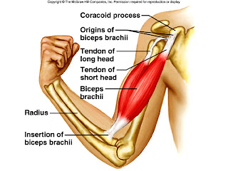
So this post is to explore the other problems with elbows – specifically "climber's elbow" – a differential elbow pain to that of tennis or golfers elbow. Less common than biceps brachii injuries but quite common in climbers.


Climber's elbow is caused by tendonitis of the brachialis muscle. The brachialis muscle lies deeper than the biceps brachii muscle and originates on the upper humerus and attaches to the ulna.

Brachialis is a true flexor of the elbow as it attaches to the ulnar (rather than attaching to the radius which rotates over the top of the ulnar during pronation and supination. N.B. Biceps brachii attaches to the radius)

Therefore, because of the broad origin on brachialis and it's only function is to flex the elbow, the brachialis can be considered the strongest elbow flexor.


This injury, if a gradual onset, is most likely to be tendonitis. If there is pain in this area of the elbow after a specific incident/fall, it could be a rupture of ligaments or muscle tendons. 

N.B. Pain in this area of the elbow, could be, as mentioned above, could be from biceps, from brachialis, or even from problems with the proximal ulna-radial ligament. Always worth getting these kind of problems checked out.

Also needing ruling out would be shoulder / wrist / finger injuries or muscular imbalances.

Palpation


To try and identify the injured structures, you can try and palpate the painful area. 
The brachialis tendon must also be palpated for tenderness during elbow flexion, as both the biceps and brachialis flex the elbow. 
The brachialis muscle and its tendon are palpated where they insert at the tuberosity of the ulna and the coronoid process of the ulna. 
Like the biceps, the distal end of the muscle and/or the insertion of the tendon would be tender with injury. 
Supination of the hand would not necessarily affect the brachialis tendon, helping to further differentiate between the two muscles.

Symptoms


  • Deep elbow pain (not superficial like tennis/golfers elbow generally is)
  • Pain on anterior (front) elbow (note, this could be due to a biceps brachii injury)
  • Swelling around the elbow or above the elbow (in the cubital fossa)
  • Inability to bend elbow comfortably
Cause

Brachialis tears normally occur during a forceful contraction or a forceful hyperextension while climbing. Complete tears are associated primarily with elbow dislocation.


Tendonitis is normally caused by strain from sudden increase in training, overuse or repetitive elbow bending or forced, excessive elbow straightening (hyper-extension).

Treatment


Full rupture = surgical intervention would be required.

For a partial tear:

  • Control inflammation in the acute phase – see POLICE Principles
  • Rest
  • Dep tissue frictions / Massage
  • It has also been suggested that traversing may irritate the brachialis, so consider limiting this.
  • If a tendonitis, the research suggests eccentric biceps curls (lowering of a weight in the curl position), however, I've found anecdotally that in the hammer position with your thumb pointing upwards works better.
  • Exercise progression from isometrics to eccentric to concentric strengthening, ensuring all are pain free, progress from isometrics once full range of movement around the elbow is achieved
Prevention


As usual, warming up is a key prevention method

Ensure adequate recovery time between climbing days


All-round strengthening of rotator cuff, biceps and brachialis can maintain a good strength all round to correct any kind of muscle imbalances.

Triceps can also need stretching/strengthening depending on the imbalance.

Stretching of lattimuss dorsi and biceps

Technique when performing pull-ups etc or when climbing i.e. making sure you lock with your lats by keeping your elbow close to your body. Try not to chicken-wing (see below)


In the bottom row of pictures, you can see the correct form for pull-ups, with the elbows tucked in
In the top row, the elbows are "winging", which makes you more prone to injury
Also note, don't snap your elbows straight when lowering from a pull-up, control the movement down.
  
Avoid:

  • pull ups with weights – your working to body strength – why do you need to be able to do pull-ups with weights?!
  • descending bachar ladder
  • down climbing campus board
  • no snapping back of elbows during climbing/pull ups

And if your wondering where I got the lovely t-shirt, go to climberagainstcancer.org 

http://www.climbersagainstcancer.org/

References

Live Strong article


Hochholzer T, Schoffl VR 2006 One Move Too Many. Lochner-Verlag, Germany

Tuesday, 4 December 2012

Surgery to the Hip

This post is to discuss common surgical interventions for the hip joint, for both hip fractures and osteoarthritis of the hip (as mentioned previously here). This will only cover those interventions performed on the bony structures, and not those to soft tissue structures that would affect the hip such as Tensor Fascia Latae (TFL) release.

There are four main surgical interventions to the hip joint, all that occur around the head and neck of the femur, and the acetabulum of the pelvis. These are:
  1. Dynamic Hip Screw (DHS)
  2. Hip resurfacing
  3. Hemiarthroplasty (Half hip replacement)
  4. Total Hip Replacement (THR)
The latter three all require the patient to adhere to precautions for a period of 12 weeks after the surgery, but there is evidence to say that these precautions are not required for hemiarthroplasties, which will be discussed later.
The latter three are also utilised as surgical interventions for osteoarthritis as well as hip fractures.


Hip Precautions (Copyright Troy Paiva)

Dynamic Hip Screw (DHS)

A DHS is usually the first option for a fracture to the neck of femur or intertrochanteric fracture.
This is where a specialist type of screw is placed through the femur and into the head of the femur.


The benefit of this surgery is no bone needs to be removed or replaced, and the hip joint does not require dislocating, therefore patient's can be fully weight bearing on the operated limb day 1 post op (as are all the other hip procedures, unless any unforeseen issues arise in theatre) and prognosis is generally good.

Hip Resurfacing

A hip resurfacing is where the head of femur and acetabulum are shaved down (literally cheese-grated - I've seen it!) and replaced with alternate fixtures. This procedure means that the head of femur is still in tact so that further replacements such as the below procedures can be implemented.


This procedure is also known as the "Birmingham Hip Resurfacing", but, unlike the DHS, the hip joint requires dislocation to get to both aspects of the joint (as do the following surgical procedures).
It is useful for the more active and younger of patients due to the hip replacement "wearing out" and so enabling further surgical interventions, as previously mentioned. It also means a lower risk of dislocation due the the similarity of size in the accompanying parts, as well as a broad range of movement and stability.

Hemiarthroplasty

A hemiarthroplasty consists of replacing only the femoral head with an artificial structure rather than the whole joint.


This means the joint is slightly more stable than its total replacement counterpart, as some of the original joint is left in place.

Total Hip Replacement

This replaces the whole joint, both the femoral component and the hip socket, the acetabulum.


The process involved in replacing the boney structures with artificial ones


The metal implant

Due to losing all the structures around the hip joint, such as the ligamentous capsule and other ligaments, and the inherent instability of an artifical joint, this replacement is a last effort and is at a higher risk of dislocation.


All the above replacement procedures can have cement or uncemented components. This doesn't impact too much and trying to explain it won't add terribly much depth to this post.

Hip Precautions

So I've already explained what the three hip precautions are, usually implemented for 12 weeks post op, and that the idea of them is to prevent hip dislocation. 
Now, the evidence behind the implementation  for total hip replacements is strong, due to the replacement of both the femoral and acetabulum aspect of the joint, then the joint is less stable and congruent, therefore dislocation risk is high.

However, with hemiarthroplasties, only the femoral head is replaced, resulting in a stabler and more congruent joint than a total replacement. Also, the surgical apporach has been changed. 

Typically, a hip will dislocate in the direction of surgical approach (Talbot et al 2002), therefore, if the posterior approach is used (as was commonly so), then the hip is going to be at a higher risk of dislocation. However, nowadays, an anterolateral approach can be used, and is preferred, as although it comes with a longer operating time, and an increase in the risk of bleeding and infections, there is a decrease in dislocations and thrombosis (in a study of 531 patients by Keene & Parker (2004)); a posterior approach places the patient at higher risk of dislocating through over-flexing because this is a routine functional position; for example, rising out of a chair. 

Studies indicate that, owing to the inherent stability of hemiarthroplasty, that is, the larger femoral head compared with the smaller femoral component in total hip arthroplasties (Baker et al 2006, Sah and Estok 2008), there is little requirement for patients to adhere to hip restrictions.
The British Orthopaedic Association and British Geriatric Society (2007) also released recommendations agreeing with the evidence already described: ‘for hemiarthroplasty introduced via an anterolateral approach … [this] should make any restrictions on hip movements unnecessary’.


However, the approach used is the decision of the person performing the operation, as both have their advantages and disadvantages, such as the soft tissue structures that are disturbed to get to the joint, as well as those mentioned above, such as risk of infection and bleeding.


How many hospitals still use hip precautions?

In a study by Fox et al (2011) in a telephone audit of all the units within the National Hip Fracture Database in England (174 units in total), 78% still used hip precautions, despite insufficient evidence. McQueen et al (2009) sent out questionnaires to members of the College of Occupational Therapists Specialist Section - Trauma and Orthopaedics, and to therapists who responded to an article in Occupational Therapy News, with a 50% response rate. The results found that 70% of therapists still implemented hip precautions following hemiarthroplasty, regardless of surgical approach.

Cost implications

From an occupational therapy point of view, hip precautions means equipment for patients to help them adhere with the precautions, and time to teach the use of this equipment and re-iterate hip precautions. In the study by Fox et al (2011), a local audit was held, and found that without hip precautions for the hemiarthroplasties, mean equipment costs were decreased from average of £49 to £37 (saving of £12 per patient), and therapist time spent with a patient was decreased by 1.5 hours (from an average of 8 hours to 6.5 hours per patient). Less equipment to order also meant that discharge delay was reduced by 0.25 days.

Hip Dislocations


Figures for hip dislocation rates vary, figures from the Cochrane review (Parker & Gurusamy 2009) reported that:
  • 33/774 hemiarthroplasties had dislocated (4.43%)
    compared to 
  • 44/333 total hip arthroplasties (13.2%). 
Other studies have results that vary, but most are similar results to that of Parker & Gurusamy (2009).
 
References


British Orthopaedic Association, British Geriatrics Society 2007 The care of patients with fragility fracture. London: British Orthopaedic Association.

Fox R, Halliday B, Barnfield S, Roxburgh J, Dunford J, Chesser TJS 2011 Hip precautions after hemiarthroplasty: what is happening in the UK and at what cost? Annals of the Royal College of Surgeons of England 93:(5) 396-397

Keene GS, Parker MJ. 1993 Hemiarthroplasty of the hip--the anterior or posterior approach? A comparison of surgical approaches. Injury 24(9):611-3.

McQueen J, Nivison C, Balance F, Fairbairn P, Clyde D, Murray E 2009 Hip precautions following hemiarthroplasty: A UK study of occupational therapists. International Journal of Therapy and Rehabilitation, 16(3):147 - 154

Parker MJ, Gurusamy KS 2009 Internal fixation versus arthroplasty for intravascular proximal femoral fractures in adults. The Cochrane Collaboration, Issue 1.

Sah AP, Estok D 2008) Dislocation rate after conversion from hip hemiarthroplasty to total hip arthroplasty. Journal of Bone and Joint Surgery, 90-A(3), 506-16.

Talbot NJ, Brown JHM, Treble NJ 2002 Early dislocation after total hip arthroplasty. Journal of Arthroplasty, 17(8), 1006-08.