Showing posts with label inflammation. Show all posts

Tuesday, 9 July 2013

Achilles Tendinopathy


So I'm going to take you back to ancient times, where Gods ruled the lands, and the Greeks invented the Achilles heel! This weeks post is going to focus on Achilles Tendinopathy.
Other common rock climbing foot issues will come in a later post.


Achilles tendon anatomy


Movements of the Achilles


Function
The Achilles tendon supports 6-15 times your body weight and provides spring like action.

Tendonitis/tendinosis/tendinopathy, what is the difference?

Tendonitis – inflammation
Tendinosis – chronic tendinopathy
Tendinopathy – a disorder of the tendons – an umbrella, catch-all term
Obviously, depending on -osis/-itis depends on the management strategies (to counter the inflammatory process or not).
The common consensus is that these conditions are a tendinosis rather than a tendonitis.

Enthesopathies
The entheses is where the tendon attaches to the bone, and an achilles tendinopathy that occurs within the first 2cm of the tendon attachment to the calcaneus is an enthesopathy.

Differential

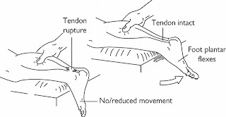
  • Rule out complete tendon rupture using the calf squeeze test. See above picture.
  • Posterior ankle impingement
  • Os trigonum syndrome
  • retrocalcaneal bursitis
  • Posterior Tibial Tendon Dysfunction
  • Haglund's deformity

However, to discuss what all of these things are would take a whole blog post unto itself, so I'm just going to leave it there!


Symptoms

  • Pain in the heel/around the tendon
  • ?swelling - if a tendinitis
  • ?heat - if a tendinitis
  • Painful to touch or on movement
  • Early morning stiffness
  • Difficulty standing on your tip-toes (/single leg stress/repetitive/hop)


Causes

Tendinopathy:
  • Repetitive strain on tendon
  • overuse
  • inappropriate footwear
  • poor technique
  • high-arched foot
  • increase intensity in training regime
  • lots of jumping
  • tight calves
  • excessive inversion or eversion

Achilles tendinopathy is more a problem for runners or walkers, but can affect climbers, especially as many climbers are mutli-sports persons.

Enthesopathies: compression of calcaneus from repeated platarflexion e.g. aggressive climbing shoes and/or dynoing etc. as climbing shoes with an aggressive heel, such as 5:10's, could cause this repeated compression on the calcaneus. (obviously, other aggressive climbing shoe are available!)


Treatment


If you suffer from the "5:10 syndrome" as mentioned above, you could try this method of editing your climbing shoes by Llanberis Resoles

NSAIDs....?
I mark this with a question mark, because it depends on your school of thought – whether it is a chronic overuse, or an inflammatory response. If you find it is swollen, then NSAID's may well help, but there is a school of thought that they may inhibit healing if used inappropriately.
There is also the dangers of overuse of NSAID's, as listed in Risks of Ibuprofen post


Acute management (see Management of Acute Injuries post) – again, may not be necessary if the tendinopathy is not an inflammatory process. e.g. if you use RSI of the wrist as an example.

GTN (Glyceryl trinitrate)
This is the same as the spray commonly used for angina, but is utilised topically – i.e. localised patch of GTN. However, the reasoning behind why it works is unclear, and commonly patients exhibit headaches and/or a rash as side effects.

Steroid
A corticosteriod injection may help, but has been shown to only have a short term pain-relieving effects, and not much else. Also, these injections may increase the risk of tendon rupture by weakening the tendon.

Surgical
A surgical intervention is rarely required for Achilles tendinopathy, and would always be a last resort. A very last resort!

There are other such treatments such as extracorporeal shockwave therapy and sclerosing injections, but they are rarely used.

The best bet is for physiotherapy, and using specific protocols, outlined below:


Physiotherapy

Mobilisations of the Achilles tendon


Ultrasound – useful if the tendinopathy is a tendonitis, as ultrasound can be used to reduce the inflammation.


Protocols/Exercises

Eccentric – Alfredson et al 1998

Two types of Eccentric Exercises will be used: (Refer to Photo A, B, and C)
  • The calf muscle is to be eccentrically loaded with the knee straight.
  • To maximize the activation of the soleus muscle, also performed with the knee bent.
  • Perform each exercise 3x with 15 repetitions.
  • Use your hand on the wall as a guide for balance
  • Begin with weight bearing load, progression to backpack weight when patient can perform the
  • exercise routine without pain or discomfort (Photo D). Advanced progression under therapist’s guidance may include resistance from weight training equipment such as a Smith machine or a squat machine.


Eccentric-concentric – Silbernagel et al 2007


Timeframe

These exercises need to be performed regularly, and for a time period of at least 3-6 months if not much longer!

Why do these work?

Effectiveness of eccentric exercises has been proved, however, there are differing theories on why it works.
Some say it affects type 1 collagen and production and, in absence of repeated aggravation, may increase tendon volume over longer term. As such, this increase the tendons tensile strength.
The repetitive stretching of the tendon with a lengthening of the muscle-tendon unit may have an impact of the capacity of the unit to effectively absorb load.

Another theory is that it changes the mechanism of pain-producing nerves by an alteration of neovascularisation (which is an increase of blood flow to an injured area, and along with this, an increase in nerve fibres, meaning increased pain), as the repetitive nature may damage these nerves and vessels.


Exercising during rehab

Due to the length of rehab being several months rehab, many patients ask “Do I have to stop exercising?” Silbernagel et al 2007 found that if the pain in your Achilles tendon does not exceed 5/10 on visual analogue pain scale (VAS) (during or after sport), then it is ok to carry on with your sport, whilst continuing with the exercise protocol.


Risk of rupture?

80% of ruptures completely asymptomatic
97% associated with underlying pathology


As always, if in doubt, always seek advice from a professional!

References


Alfredson H, Cook J 2007 A treatment algorithm for managing Achilles tendinopathy: new treatment options Br J Sports Med. 41(4): 211–216.

Silbernagel KG, Thomeé R, Eriksson BI, Karlsson J 2007 Continued Sports Activity, Using a Pain-Monitoring Model, During Rehabilitation in Patients With Achilles Tendinopathy: A Randomized Controlled Study. American Journal of Sports Medicine 35(6): 897-906

Alfredson H, PietilÀ T, Jonsson P, Lorentzon R 1998 Heavy-Load Eccentric Calf Muscle Training For Chronic Achilles Tendinosis American Journal of Sports Medicine 26(3): 360-366

Thursday, 13 June 2013

"Climber's elbow" - Brachialis Tendonitis

So this post is to explore the other problems with elbows – specifically "climber's elbow" – a differential elbow pain to that of tennis or golfers elbow. Less common than biceps brachii injuries but quite common in climbers.


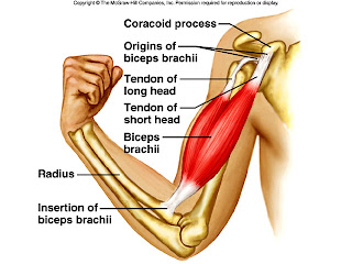
Climber's elbow is caused by tendonitis of the brachialis muscle. The brachialis muscle lies deeper than the biceps brachii muscle and originates on the upper humerus and attaches to the ulna.

Brachialis is a true flexor of the elbow as it attaches to the ulnar (rather than attaching to the radius which rotates over the top of the ulnar during pronation and supination. N.B. Biceps brachii attaches to the radius)

Therefore, because of the broad origin on brachialis and it's only function is to flex the elbow, the brachialis can be considered the strongest elbow flexor.


This injury, if a gradual onset, is most likely to be tendonitis. If there is pain in this area of the elbow after a specific incident/fall, it could be a rupture of ligaments or muscle tendons. 

N.B. Pain in this area of the elbow, could be, as mentioned above, could be from biceps, from brachialis, or even from problems with the proximal ulna-radial ligament. Always worth getting these kind of problems checked out.

Also needing ruling out would be shoulder / wrist / finger injuries or muscular imbalances.

Palpation


To try and identify the injured structures, you can try and palpate the painful area. 
The brachialis tendon must also be palpated for tenderness during elbow flexion, as both the biceps and brachialis flex the elbow. 
The brachialis muscle and its tendon are palpated where they insert at the tuberosity of the ulna and the coronoid process of the ulna. 
Like the biceps, the distal end of the muscle and/or the insertion of the tendon would be tender with injury. 
Supination of the hand would not necessarily affect the brachialis tendon, helping to further differentiate between the two muscles.

Symptoms


  • Deep elbow pain (not superficial like tennis/golfers elbow generally is)
  • Pain on anterior (front) elbow (note, this could be due to a biceps brachii injury)
  • Swelling around the elbow or above the elbow (in the cubital fossa)
  • Inability to bend elbow comfortably
Cause

Brachialis tears normally occur during a forceful contraction or a forceful hyperextension while climbing. Complete tears are associated primarily with elbow dislocation.


Tendonitis is normally caused by strain from sudden increase in training, overuse or repetitive elbow bending or forced, excessive elbow straightening (hyper-extension).

Treatment


Full rupture = surgical intervention would be required.

For a partial tear:

  • Control inflammation in the acute phase – see POLICE Principles
  • Rest
  • Dep tissue frictions / Massage
  • It has also been suggested that traversing may irritate the brachialis, so consider limiting this.
  • If a tendonitis, the research suggests eccentric biceps curls (lowering of a weight in the curl position), however, I've found anecdotally that in the hammer position with your thumb pointing upwards works better.
  • Exercise progression from isometrics to eccentric to concentric strengthening, ensuring all are pain free, progress from isometrics once full range of movement around the elbow is achieved
Prevention


As usual, warming up is a key prevention method

Ensure adequate recovery time between climbing days


All-round strengthening of rotator cuff, biceps and brachialis can maintain a good strength all round to correct any kind of muscle imbalances.

Triceps can also need stretching/strengthening depending on the imbalance.

Stretching of lattimuss dorsi and biceps

Technique when performing pull-ups etc or when climbing i.e. making sure you lock with your lats by keeping your elbow close to your body. Try not to chicken-wing (see below)


In the bottom row of pictures, you can see the correct form for pull-ups, with the elbows tucked in
In the top row, the elbows are "winging", which makes you more prone to injury
Also note, don't snap your elbows straight when lowering from a pull-up, control the movement down.
  
Avoid:

  • pull ups with weights – your working to body strength – why do you need to be able to do pull-ups with weights?!
  • descending bachar ladder
  • down climbing campus board
  • no snapping back of elbows during climbing/pull ups

And if your wondering where I got the lovely t-shirt, go to climberagainstcancer.org 

http://www.climbersagainstcancer.org/

References

Live Strong article


Hochholzer T, Schoffl VR 2006 One Move Too Many. Lochner-Verlag, Germany

Thursday, 9 May 2013

The Risks of Ibuprofen

So, you have some swelling, after an injury, or maybe it's a chronic condition? So you pop some ibuprofen or two?

After a while, you realise the ibuprofen is no longer having the same effect, so you take some more to reach the same level of pain relief?

Before you know it, you've been taking it for weeks/months/years.


Sound familiar?


I hope not.


Non-steriodal anti-inflammatories drugs (NSAIDs for short) have there place and can be relevant when taking appropriately. However, I wanted just to discuss some recent evidence that was brought to my attention about the dangers of NSAIDs (not to scare anyone, but just to inform you).


It has been widely documented that NSAIDs can cause kidney damage (Murray & Brater 1997) due to all NSAIDs reduce blood flow to the kidneys. As a result 2 things can occur: elevation of blood pressure; and, more dangerous, the risk of acute renal failure/acute tubular necrossis.


But other studies have now shown the they may also cause damage to the intestines, and to bone healing.


Bone Healing:


A recent systematic analysis of studies looking at affects on bone healing caused by NSAID's by Pountos et al (2012), and found that, although there is an absence of robust clinical trials, that due to the scientific knowledge of the interference at a cellular level caused by NSAIDs, that they should be viewed as a risk factor to bone healing.

The idea is, due to the anti-inflammatory effect of NSAIDs, that it reduces the amount of certain chemicals in the cells that aid healing, as they are induced by the inflammation process.

These chemicals are thought to enlist osteoblasts which are responsible for bone formation.


Osteoblasts via microscope


Intestinal damage:


It has already been tested that you get some marginal intestinal damage when exercising (due to functions such as digestion become a luxury when exercising, as the blood that normally supplies the small intestine is instead diverted to the muscles. Due to this lack of blood to the intestines, some of the cells lining the intestines are traumatized and start to leak.)


However, it has been shown in a recent study by Van Wijck et al (2012) that NSAIDs, specifically ibuprofen, can aggravate this phenomena. To what extent, I'm not too sure, but the result was a significant one.

It is worth noting that this was performed on healthy males performing an aerobic exercise (cycling), and only 9 subjects were used, and is only a short term effect. The long term effects and consequences are not yet clear.


So, there you have it. I'm no pharmacist or chemist, but I thought I'd share with you what I'd read. Draw whatever conclusions you wish to from this, but it's worth bearing in mind.


References


Murray MD, Brater DC 1997 Effects of NSAIDs on the kidney. Prog Drug Res. 49:155-71.


ReynoldsG 2012 For Athletes, Risks From Ibuprofen Use. New York Times Blog


VanWijck K, Lenaerts K, Van Bijnen AA, Boonen B, Van Loon LJ, Dejong CH,Buurman WA 2012 Aggravation of exercise-induced intestinal injury byIbuprofen in athletes. Med Sci Sports Exerc 44(12): 2257-2262


PountosI,GeorgouliT, Calori GM, Giannoudis PV 2102 Do Nonsteroidal Anti-InflammatoryDrugs Affect Bone Healing? A Critical Analysis. Scientific WorldJournal 606404


Friday, 19 April 2013

Physiological response to injury and hot/cold

So, this post originally started off as a post to review the evidence behind contrast baths, however, I realised it would be best to explain the physiological effect of hot and cold on the body, and discuss healing times while I'm at it, due to the overlap.

So, here it goes, I will begin with healing times and process.

The healing process consists of 3 different phases, and these are:
  1. Inflammation (0-48 hours)
  2. Proliferation (5 days – 4 weeks)
  3. Maturation (4 weeks – 2 years +)

Now, I've put the time frames up here as a guide. A popular misconception is that these time frames are distinct and seperate, as shown below:


When actually, each of these areas overlap, as one area is winding down, the next is building up it's response, as shown below:

Now, here I will discuss what each area does physiologically, in as brief and simplest way possible:

Inflammation

We are all familiar with this process. This is when the injured area, swells, become red in colour, with associated warmth, pain, and loss of function. In this stage, the initial bridging of collagen fibres begins.
The normal response in this stage is to apply the POLICE principles.

Now, I must point, at this stage, we all panic about swelling, and try our best to get rid of it. Swelling is a natural part of the healing process and we should let it take it's course, as the swelling is needed to get fresh nutrients to the area, and to remove cellular debris.
Chronic swelling, so swelling that is still present maybe a week after the injury is not necessarily good, as this will start to impact on the optimisation of the healing process, and this is where we would want to remove excess inflammation.
Swelling upon exercise of the injured area, at this stage, would still be normal.

Proliferation

This stage shows the signs of inflammation beginning to decrease, and pain levels decreasing too.
Collagen fibres are being laid down at the injured site at the peak rate at this stage and will progress to orientate themselves in line with imposed stresses.

Maturation

This stage should so no signs of inflammation and a huge decrease in pain, with pain only at end range. This stage should focus on restoring function and strength, and optimising collagen alignment.
Collagen fibre deposits peak around 3 months post injury, but will continue to lay down up to 2 years after an inujry (depending on the severity of the injury).
Maturation is more focussed on return to normal, with excess collagen fibres removal, optimisation of the collagen matrix to accommodate imposed stresses, and return to normality of the vascular supply.


This video discusses the three stages of healing in much more
detail, if that interests you, and you can stand the accent!

N.B. Pain is not synonymous with healing. Pain levels will decrease quicker than structural integrity of the injured site improves, so the risk of re-injury is still high, even if the pain has subsided.

This graph shows that after the injury, the pain levels drop
below the threshold quicker than the tendon recovery

It is also worth mentioning a few stats on the temperature of tissues.

Optimal tissue temperatures:
resting = 36.5ÂșC – 37.2ÂșC,
aim when exercising = 38ÂșC – 40ÂșC (as tendons exhibit plastic deformation at 39ÂșC, collagen at 40ÂșC)

Hot and Cold response

So, now I'll move onto the effect of hot and cold physiologically:

Hot and cold can increase or decrease symptoms of inflammation, and here's how:

Cold
Glenn ice climbing in cold conditions!
Copyright Glenn Manifold

Symptom
Response
How?
Additional info:
Pain
Reduces nerve conduction velocity and increases the firing threshold
Considerations
Rewarming after icing can take 3-4 hours
With a muscle at 4cm depth from skin surface will have a 4ÂșC fall in 1hr

Contraindications: Raynauds, Cold hypersensitivity, Open wound, Sensory defect, heart disease

Precautions: Cardiac disease/hypertension, pelvis/groin area (bone marrow production/blood cells), left shoulder+neck area (proximity to heart)

Spasm
Automatic protective response, prevents increase demand for O2
Metabolism
Reduces need for O2 in surrounding tissue so less damage via hypoxia and reduces total debris. Less O2 means less secondary damage to other tissues
Blood flow
Vasoconstriction of the blood vessels
Inflammation

Oedema
By vasoconstriction of blood vessels and increasing blood viscocity
Tissue extensibility


Heat
The author bouldering in sunny,
warm Fountainebleau
Symptom
Response
How?
Additional info:
Pain
By reducing the nerve conduction velocity and increasing the firing threshold
Precautions/contraindications: Diabetes mellitus Multiple sclerosis Peripheral vascular disease

Spinal cord injuries Rheumatoid disease


Possible side effects: skin burns
Spasm
Automatic protective response, prevents increase demand for O2
Metabolism
More energy to cells, increasing there productivity, plus increase in blood flow meaning more oxygen to area
Blood flow
Vasodilation of the blood vessels
Inflammation

Oedema
By vasodilation of blood vessels and decreasing blood viscocity
Tissue extensibility


I hope this post has given you a little more understanding around the healing process and the physiological effects of hot and cold, and hopefully this will aid understanding for future posts, such as the evidence behind contrast baths, that I'm currently working on.